Patient-specific cardiac digital twin · research preview

Rehearse the MitraClip before you touch the patient.

One routine cardiac CT becomes a beating, patient-specific twin of the heart and mitral valve. Place a virtual clip and the whole valve re-balances into a new equilibrium — the leak falls, forward flow recovers, and the trade-off shows itself. Live, on the patient's own anatomy.

Research use only · not a medical device · a model, not a clinical prediction
moving the clip · the valve re-balances
Move the clip · watch the valve respond
Leak
↓ better
Forward output
↑ better
Gradient
watch
01 · Why CT

Echo reads the valve. CT lets you compute it.

Echo stays the workhorse for grading the leak and guiding the table in real time. But a digital twin needs a substrate it can solve on — an isotropic, full-cycle 3D volume that comes out the same every run, independent of the operator and the acoustic window. One routine cardiac CT gives you that.

Echo today
CT digital twin
Dimensionality
2D planes you assemble in your head; the 3D lives in the operator's mind.
A true 3D volume — the same in every direction, nothing to reconstruct by eye.
Whole cycle
Real-time and functional, but sampled plane by plane as the probe moves.
Every phase of the beat in one shared frame — chambers, both valves and blood already aligned.
Reproducibility
Operator- and load-dependent; a second read can land somewhere else.
Same volume, same result, every run — independent of who scanned it.
Computable substrate
Hard to turn a live acoustic read into a solvable mechanical model.
A geometry you can mesh, free as physics objects, and solve to equilibrium.
Operator dependence
Bound to the acoustic window and the hand at the probe.
No manual tracing in the loop — the volume goes in, the twin comes out.

Echo keeps its job — the twin doesn't replace the read. It adds a substrate you can compute on.

02 · From CT to twin

From one CT to an operatable twin.

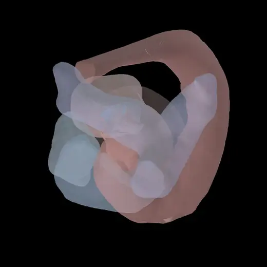
Not a picture of the heart — a model you take to the table. The chambers and both valves reconstruct into a beating twin you can scrub, with the patient's own mitral leaflets moving through every frame.

  • A beating whole heart, reconstructed from the scan you already order
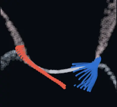
  • The patient's own leaflets — coapting as the valve shuts, opening as it fills
  • Calcium kept separate, never fused into healthy leaflet tissue
operatable — taken to the table virtually
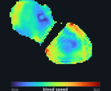
live twin · beating4D · one cardiac cycle
beating reconstructed heart twin
03 · Flow simulator

See the leak the valve can't stop.

With the valve moving, the blood moves with it. When it shuts, a leak the valve can't stop drives back toward the atrium — exactly what the clip is meant to correct. Two views, side by side, running through the beat.

long-axis cutthe leaflet gap
long-axis cut through the valve across the beat
blood speedthe regurgitant jet
blood-speed flow field across the beat
The flow is shown qualitatively — the point is the leak, and how placing the clip re-balances the valve to cut it. The hero shows that trade-off live; the twin reads it back on the patient's own anatomy.
The future · in build

Not open yet. Be first when it is.

We're building this toward the structural-heart team. Leave your email and we'll show you the twin the moment it's ready — no access today, just first in line.

Get notified →
05 · Under the hood

Three models. One physics-true valve.

The clip result is mechanics, not a lookup. Three models stack: we recover the patient's own moving leaflets from CT, drive blood through them off the patient's own beat, then bind geometry and flow with a physics solve that recovers what CT never saw.

Pillar 01

Leaflet reconstruction model

Recovers the patient's own moving leaflets from a single cardiac CT — both leaflets tracked as one sheet across the whole beat, calcium separated from healthy tissue. The valve is thin, fast and barely resolved on CT, so most pipelines never see it move. This one is purpose-built to track it frame by frame.

the patient's leaflets, moving
Pillar 02

Hemodynamic simulation

Turns the moving geometry into blood. It resolves the transmitral inflow, the regurgitant jet and the diastolic gradient — driven by the patient's own beat, not a generic waveform. Pre-clip, it shows a valve that can't seal: much of each beat thrown backward instead of forward.

their beat becomes flow
Pillar 03 · the binder

Convergence-equilibrium calibration

The reconstruction is a recording of where the leaflets actually were; here we free them as physics objects and let them converge back onto that recording under a solve. That recovers the hidden chordae tension and tissue stiffness and yields a true mechanical equilibrium. Add a clip and it's just one new constraint — the whole valve re-balances into the double orifice.

geometry meets physics → equilibrium
Built on the established edge-to-edge repair — COAPT / EVEREST
Aligned with published CT-based modeling of post-clip hemodynamics
06 · The platform · your experience

Upload a CT. We build the rest.

No segmentation, no meshing, no scripting. Drop in the scan you already order and the platform does the work — then hands back what you need to decide.

Step 01

Upload your CT

The scan you already order. Drag, drop, done — no special protocol.

Step 02

See the twin

An operable, beating heart and valve, reconstructed for you to scrub.

Step 03

Simulate the repair

Place a virtual clip and watch the valve re-balance into a new equilibrium.

Step 04

See the outcome

Where the leak lands and what it costs — read back, side by side.

We hand you the read, not just a prettier picture.

Get notified · the future

Be the first to see it.

It's not open yet. Leave your details and we'll reach out the moment the twin is ready for you to try — and you can tell us what you'd want it to answer.

  • 🫀First in lineWe'll show you the twin before it opens widely — your own anatomy, walked through with you.
  • 📐Built with your inputTell us what the twin must answer to earn a place in the heart team — and where it isn't yet validated.
  • ✉️No access pitch, no spamJust a heads-up when it's ready, from a real cardiology-aware human.
Research use only — not a medical device, not for clinical decision-making, diagnosis or treatment, and not cleared by any regulatory authority. Nothing here is available for clinical use today; this is an early research preview in active development.
Get notified →
research preview · coming soon